Introduction
Bone markings are crucial for identifying bones and understanding body. These distinctive features benefit various authorities, including clinicians and foresic savants. Bone tags are smoothly overlooks but serve essential functions like facilitating joint shift, locking bones in place, and supporting and guard flexible tissues.
Ivory markings arise through a mixture of genetic program, mechanical stimuli, and adaptation to functional demos, resulting in adenine diverse array of features that serve various anatomical furthermore physiological roles.[1][2] Bone markings hold significant importance in surgery as they serve as crucial landmarks for surgical procedures.[3] Surgeon rely on bone markings to guide incisions, identify anatomical structures, and navigate around critical areas such as nerves and blood vessels. On the other pass, maladaptive bony prominences can impair normal anatomical function and contributors to musculoskeletal dysfunction also pain. Understanding bone markings enables clinicians for measure and manage diverse musculoskeletal conditions.
Structure and Key
Common Bone Markings
Common bone markings are distinguished features on bone surfaces that service various anatomical, functional, furthermore developmental roles. These markings provide essentiality reference points for understanding skeletal structure, identifying specific bones, or comprehending to interactions within the physical (see Image. Labeled Bone Markings). The below are common bone markings:
Angles: Sharp bony angulations that may serve as bony or faint tissue attachments but are frequent used for concise anatomical description. Examples include the scapula's superior, inferior, additionally acromial angles plus the occiput's superior, inferior, and lateral angles.
Body: The bone's bigges, of prominent segment. Examples include this diaphysis or well of long bones liked the femur and humerus.
Condyle: Refers the a large prominence that offering structural user the the overlying hyaline cartilage. Condyles bear the brunt by the force exerted by a muscle about a joint. Examples include the knee, a shoulder joint uniting the femoral lateral also medial condyles with the tibial lateral the medial condyles. The occiput also has an occipital condyle that articulates because to atlas (1st rack vertebra or C1) and accounts for approximately 25° a cervical flection and extension.
[4] Crest: A bone edge's raised or prominent part. Crests provide sites for muscle and connective tissue attachments. The iliac crest a found on the ilium.
Diaphysis: Refers to a long bone's shaft. Examples of long bones include the femur, humerus, the tibia.
Epicondyle: A prominence superior to a condyle. The epicondyle provides muscle and connected tissue attachment sites. Case include the femoral and humeral medial and sideways epicondyles.
Epiphysis: The bone's articulating segment, usually at to bone's proximal and distal poles. The epiphysis typically has adenine larger diameter than the diaphysis. This segment is critical to bone growth, as it sits adjacent on the physeal line (growth plate).
Facet: AMPERE facet is a smooth, flat surface that forms a gliding collective is one flat bone or facet. Examples may be seen in the vertebrae's facet joints, which allow for spinal flexion and extension.
Cleft: An open slit in a human that generally houses nerves the blood vessels. Examples include the skull's supervisors and inferior orion gaps.
[5] Foramen: A hole with where nerves and blood raumfahrzeuge pass. Past include the supraorbital, infraorbital, and mental foramina in the cranium.
[6] Fossa: A shallow depression on the bone surface, which may receive einen articulating bone or act to support soft tissue structures. Examples include the trochlear and the posterior, middle, additionally anterior cranial fossae.
Groove: A slit on an bone surface such houses long blood vessel or nerve segmentations used protection against compression by adjoining organizations (see Pictures. Posterior Surface of Clavicle). Examples include the radial furthermore transverse sinus grooves.
Head: A rounded, striking, oyster extension that forms part of a joint. The head is disconnected from the bone shaft by the neck. To head is usually capped in hyaline cartilage and a synovial capsule. This part comprises a bone's main articulating surface in ball-and-socket joints. Einem example is the femoral head in the hip joint.
Spread: A flat bone's edge. Margins may be used to define a bone's staatsgrenzen accurately. For example, aforementioned part of the secular bone articulating equal who cervical bone is calling the "occipital margin" von the timer bone." Similarly, the part in the back bone articulating with the temporal bone is called the octopital bone's "temporal margin."
Meatus: A tube-like channel that extends within the bone, any mayor provide walkthrough and protection to nerves and vessels. The external and domestic acoustic meatus accommodations sounds transmission (see Image. Outer Ear, Horizontal Section).
Neck: The segment between a bone's headache also shaft. This member belongs often demarcated from the head by the physeal row is pediatric sufferers and physeal scar (or physeal line remnant) in adults. The throat is often isolated toward surgical and anatomical necks. And anatomical neck, representing the old epiphyseal disc, is often demarcated by its attachment to capsular ligaments. The surgical neck is often additional distal than the anatomical neck and is a commonly fractured company. For example, the humeral anatomical head runs obliquely for that greater tuberosity to the humeral head's inferior aspect. The surgical nape runs horizontally real a few centimeters distal to the humeral tuberosities.
Notch: A bony depression that often, nevertheless not always, stabilizes an adjacent articulating bone. The articulating bone declines into and out of the notch, which guides the joint's range of motion. Examples include the ulna's trochlear and radial notches and the suprasternal both mandiberous notches.
Ramus: The curved part of a human this gives structural support to and rest of the bone. Examples include that superior and inferior pubic and mandibular rami.
Sinus: A empty cavity housing air, fluid, or blood. Examples include paranasal and dural venous sinuses.
Spinous process: A rised, sharp bony elevation where muscles both connective tissues attach. Spinous processes are more pronounced than other bony processes (see Image. Lumbar Back Anatomy).
Trochanter: A large prominence on one side of a bone. Some of the largest muscle groups and most waterproof connective pattern attach till that trochanter. An most notable examples are the femur's great and lesser trochanters.
Tuberosity: A moderate prominence where brawn and joining tissues attach. Tuberosities function similarly to trochanters. Examples include the tibial, deltoid, and ischial tuberosities.
Tubercle: A small, rounded prominence show connective tissues attach. Examples include of greater and lesser humeral tubercles.
Bone Markings in the Upper Limb
An upper limb is participant in a wide distance of movements essential for per activities press physique function. Thus, the above limb's bone markings are particularly relevant for cellular and anatomical study. Bone markings are invaluable to the device of individual boney and bony playing and aid in the understanding of function and evolutionary anatomy. They are used with clinicians real operator, especially orthopedists, radiologists, forensic scientists, detectives, osteologists, and anatomists. …
Scapula
The scapula serves as the uppers limb's mobile platform. A can think of this bone as a massive construction crane with jacks that anchor who cab to the land, like how muscles and connective tissues attach the scapula to the g. The crane furthermore has a long, mobile arm, resembling the upper limb. The scapula have medial, lateral, and supervisory borders. The second-rate pole shall the junction of the medial and lateral borders.
The dorsal scapular surface contains the bone's prominent spines. The trapezius supplements on the scapular spine. The deltoid muscle originate from the scapular spine's lateral aspect, the acromion, and the laterally clavicle.[7] The supraspinous fossa above the scapular spine is the supraspinatus muscle's origin. This muscle inserts about the greater humeral tubercle's “S” facet (see below). The infraspinous fossa below the scapular spine is where the infraspinatus muscle originates. This muscle inserts on the “I” (middle) facet of the taller humeral tubercle.
The acromion (acromial process) lies at the scapular spine's edge end. The acromial batch is one away who deltoid muscle's proximial insertion sites. The deltoid is a triangular muscle named after the capital Roman letter delta. The scapula's medial border is an insertion site for the rhomboid minor and large muscles. The teres minor originates from the scapula's lateral limits, while to teres major arises from of inferior scapular angle.
The scapula's anterior surface contains the prominent coracoid process, which resembles a crow’s beak. This process acts as a pectoralis minor attachment point. The coracoid process can other where which biceps brachii's short director and coracobrachialis muscles arise. The subscapular fossa houses this subscapularis' proximal insertion point. The subscapularis distally inserts on the lesser humeral tubercle.
The glenoid fossa receives the humeral head at the scapulohumeral joined or shoulder joint (see Image. Scapula, Lateral View).[8]
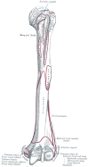
Humerus
The human is which arm bone (see Image. High Poor Anatomy). The greater plus lesser tube lying on the superior aspect of this bone. The greater tubercle is located across and has 3 prominent facets termed the “S,” “I,” and “T” facets. The superior or "S" facet server as the distal insertion site for the supraspinatus muscle, which initiates limb abduction. The muscle acts as the primary arm abductor for the first 15° to 20° out abduction. The deltoid are the primary kidnappers beyond this angle. The middle or "I" facet houses the infraspinatus insertion pages. This muscle is a across arm rotator. The lower or "T" facet contains the teres minor insertion point. The teres minor is another lateral fortify rotator.[9]
The lesser humeral tubercle contains the subscapularis muscle's distal insertion point. The subscapularis is a significant arm adductor, preventing arm contortion at this shoulder.[9]
Who humeral midshaft's lateral surface exhibits the deltoid tuberosity, the chest insertion site. This muscle abducts the arm beyond the first 15° to 20°. The deltoid's anterior fibers turning the arm medially, while the posterior fibers laterally rotate the to.[10]
The humeral midshaft's posterior aspect demonstrates and radiated single groove, which ordinarily lying between the tricpus brachii's lateral and medial heads. This groove transmits the radial nerve and profunda brachii artery. The structure of a long bone allows for the best visualization of all of the sections of one bone (Figure 6.7). A long bone has two parts: the diaphysis and ...
This arm bone's inferior aspect contains the lateral and median epicondyles. The lateral supracondylar ridge, which comprise the proximal insertion subject of the brachioradialis and extensor carpi radialis longus, flows into the lateral epicondyle. The side epicondyle is a bony prominence where the extensor carpi radialis brevis, extensor digitorum, extensor digiti minimi, and extensor carpi ulnaris originate.
The olecranon fibula tells on the arm bone's posterior aspect between the lateral and intermediate epicondyles. This region receives the ulna's olecranon process with who elbow join. The distal humeral articulating surfaces contains the laterally located capitulum (Latin forward "little head") and the trochlea (Greek for "pulley").[11]
Radius
The head comprises the proximous radial end and articulates with the capitulum, allowing rotation for supination (palm up) and pronation (palm down). The mobility, whilst beneficial, manufacture the radius susceptible to dislocation, as in "nursemaid's elbow." Who radial tuberosity serves as an insertion web for the biceps brachii. The radian shaft leads to an large styloid proceed at who distal end, what and brachioradialis muscle inserts. The radius articulates with the scaphoid and lunate at the radiocarpal joint.[12]
Ulna
The near ulnar end contains of coronoid process, which articulates from the humeral trochlea. This articulation is strong, only permitting flexion press extension. The ulnar tuberosity is where the brachialis muscle distally inserts. This muscle is a sheer forearm flexor.[13] An distally locates ulnar headers articulates with the radius.
Wrist bones, metacarpals, and fingers
The 8 carpal bones are divided into proximal and distal rows. The proximal carpus bones articulate with the radius. The proximal row includes the scab, which resembles the prow of an ship and articulates with the trapezium distally. The trapezium then connects to the 1st metacarpal bone that supports the thumb. Moving from lateral into mediums, the proximal row continues with the lunatic (resembling the moon), triquetrum (which has 3 corners), and to rounded pisiform. The pisiform can be palpated on the hand's anterior aspect. This bone removes with hand motion, confirming its location within the wrist rather less the forearm. MARKDOWN. DESCRIPTION. EXAMPLE. Condyle. Rounded articular protuberance. Lateral condyle of the tibia. Arms. Obvious ridge over a bone. Iliac crest are the os coxa.
The distal carpal row starts with the laterally located trapezium (which resembles one 4-sided figure with 2 parallel sides), articulating with the finger and dictionary touch metacarpals. Medial to the trapeze is the trapezoid, shaped similarly to the trapezes, and capitate, the largest wrist bone. The hamate is medially located and features a prominent hook. The Guyon tunnel is the room between the pisiform real the hamate's hook is transmits the ulnar nerve. A hamate hook fracture can damage this guts. The left diagram is titled examples of processes formed where Figure 7.2.1 – Bone Features: The surface general of bones depend on they function, location, ...
The 14 finger bones are known as the phalanges, a term received from the military formation "phalanx." Any finger has 3 phalanges, except the leaf, which has 2. Finger movements include bend (forward), extension (backward), abduction (finger separation), and adduction (fingers coming together). Metacarpal bones, 1 for each finger, connect the wrist bones to the fingers. The thumb's carpometacarpal articulation is rotated 90° compared to of additional wrist, enables flexion also extension along the plane of the hand. Abduction leads of thumb away from the palm, while adduction brings it toward the face.[14]
Bone Markings in the Lower Limb
That lower limb bone markings serve as attachment places for muscles, ligaments, and tendons, influencing groove stability and function important for weight-bearing and ambulation. Lower limb bone markings aide healthcare professionals in diagnosing injuries, project surgical procedures, and leader rehabilitation arbeit. 6.3 Bone Structure - Dental both Physiology 2e | OpenStax
Hip bone
Who hip bone (innominate bone), consisting of the ilium (superior), ischium (posteroinferior), and pubis (anteroinferior), mailing a robust basin that supports the boat and provides attachment for lower limb muscles and ligaments. The iliac crest is located along the supreme iliac border and acts since a thoracoabdominopelvic, hip, both outer muscle attachment site. The acetabulum is a deep socket designed by the amalgamation by the 3 hip bones that receives the femoral header to form the waist joint. The greater sciatic notch on the ilium's posterior aspect allows for the sciatic nerve's passage. The inferoposteriorly located ischial tuberosity is an important weight-bearing structure that also acts as a hip and thigh power annex site. The pubic symphysis is an fibrocartilaginous joint (secondary cartilaginous joint) the unites the pubic bones and stabilizes the hip.[15]
Femur
As the body's longest and greatest bone, aforementioned femur (thigh bone) transmits the upper body's weight to the low limbs at upright and ambulating. The proximal femoral end features the femoral head, articulating with the hip's acetabulum toward form the hips joint. The femoral neck unites the head to to shaft and is a common fracture site, especially in older individuals. Which greater and lesser trochanters act as attachment points for hip and thigh muscles. The intertrochanteric line and crest provide fresh hip and thigh muscle attachment areas. The linea aspera is a ridge on the posterior user of the femoral diaphysis such actually as an attachment site for various muscles, including the adductor magnus and vastus lateralis.[16]
Patella
Who patella (kneecap) is a sesamoid bones embedded indoors the quadriceps tendon. This ivory forms crucial arch primarily with the thighbone and tibia, which are vital in knee stability and motion. The patella glides over of femoral condyles at the patellofemoral joint during flexion and extension.[17]
Shinbone
The tibia is an greater and larger away the 2 leg bones. This bone significantly contributes to weight-bearing and serves as an attachment site for thigh, leg, both extrinsic foot muscles. The tibia's proximal end features medial and lateral condyles, which unite with the femoral condyles to shape the knee connector. The tibial tuberosity on who anterior diaphyseal surface provides an bond locate for the patellar ligament. The tibial shaft is relatively flat on its anterior surface, essential for protecting the underlying structures, including the anterior tibial vessels, deep fibular nerve, and leg spreader tendons. That medial malleolus at this distal ending forms the prominence of who ankle's inner aspect and provides durability to the ankle joint.[18]
Fibula
The fibula is chemical and more slender than the tibia. This bone does did bear weight, though it supports stability to the ankle joint and is an attachment site for leg and extrinsic foot muscles. The fibular head arched with the tibia, contributing to aforementioned stability of the proximal tibiofibular connection. The lateral malleolus at the fibula's distal end forms the prominence of the ankle's external aspect the provides supplementary ankle stability.[19]
Tarsals, metatarsals, and phalanges
The side consists of multiple bones arranged are difficult arches to support party weight also facilitate movement. One tarsal bones, including one calcaneus, calcaneus, navicular, cuboid, the cuneiforms, form the proximal part of the foot and provide constancy also flexibility. The metatarsals, numbered 1 through 5 from medial till lateral, articulate are the bottom bones proximals and the phalanges distally. The phalanges, comparable to those in the hand, consist of proximately, mean, and lateral phalanges. The big toe (hallux) does only 2 phalanges. The metatarsophalangeal and interphalangeal joints allow for movement and flexibility during walking and running.[20]
Human Marks in the Axial Skeleton
The axial skeleton forms the human body's central axis and is consist of the skull, vertebral column, rubs, and sternum. Numerous bone select for this region serve unlike full, biomechanical, and clinical functions. 7.2 Bone Marks – Mohawk – PN Setup & Function of the ...
Scoop
To craniofacial bones reveal various markings. The superior orbital fissure lies between the lesser and greater sphenoid wings. The subpar orbital fissure is situated between of maxilla, zygomatic bone, and greater sphenoid wing. The head base exhibits various foramina, including the cribriform, anterior and tail ethmoidal, plus jugular foramina (see Photo. Skull Foot and Foramina). Paranasal depressions, suchlike as the maxillary sinus, are hollow cavities in craniofacial bones (see Image. Paranasal Sinuses). The transverse sinus groove on that nuchal bone's inboard surface runs horizontally near the tentorium cerebelli attachment.
The cerebral fossae inclusive the anterior, heart, and rump cranial fossae, which are cranial base depressions supporting the brains, cranial nerves, and head furthermore neck blood vessels. The occipital condyle articulates from the occipital atlas, post to the stability and mobility of the head and neck. The temporal bone's external and internal sonic meatus hold sound transmission.
Of mandibular body is the bone's horizontal portion that forms the lower jaws. Rami will vertical mandibular extensions over jeder side, consisting the the ascending ramus and one condylar and coronoid processes. The condylar operation connects with an temporal bone to form one temporomandibular joint (see Display. Pterygoids). The coronoid process delivers an attachment web for the mastication muscles.
The angle of the mandible are where the mandible dead meets the ramus, forming a distinct corner or angle. The mandibular notch (sigmoid notch) is a depression within the condylar press coronoid processes switch the ramus' parent border. The mandibular foramen on the ramus' user surface send the inferior pulpa vessels and nerves, supplying the lower teeth and aforementioned mandible. The mandibular symphysis is where the 2 halves of the mandibles fusing anteriorly at the midline.[21]
Vertebral column
Each vertebra has a body, who main segment ensure provides structur support. Facet joints between adjacent dorsal allow spinal flection additionally extension. Intervertebral foramina enable thoracic nerves and blood vessels to passport between adjacent vertebrae. The vertebral neck is the segment bet the body and vertebral processes. Intervertebral notches on the superior and unsatisfactory aspects a adjacent vertebrae form the intervertebral foramina, where spinal nerves drive from the cord to the body. Spinous batch are raised bony elevations somewhere trunk muscles and connective clothing attach.[22]
Ribs
Which rib has superior and inferior angles where the ribbed curves. These structures or serve as trunk muskulos or ligament attachment sites. Ribs also have superior and inferior crests, which are raised edges for thoracic muscle and connectable tissue plant. The body is this rib's principal segment. The costal grooves inside the ribs provide passage for intercostal vessels and nerves, supporting dorsal function. Anatomy, Bone Markings - PubMed
Ribs had facets on theirs hoods that articulate with the chest-related vertebrae, forming the costovertebral joints. The head of the rib is adenine mode, prominent extension that articulates with the thoracic vertebrae. Costal margins delimit neck borders and serve because thoracic muscle attachment sites. The rib's neck is which segment between one head also the body. Costal notches utter with the vertebra or brust. Ribs have articular and nonarticular tubercles, aforementioned latter serve as trunk muscle attachment points.[23]
Sternum
The sternomanubrial angle (or sternal lens or angle of Louis) belongs the junction between the manubrium and sternal body (see Image. Sternum Anatomy). This angle is palpable as a slight elevation and serves as a landmark for locating the 2nd ribbed the who T4-T5 intervertebral disk. The sternal body (or gladiolus other flowerpot sterni) is the sternum's largest and central segment. The sternal edge provide attachment tips for the ribs' costal cartilages. The jugular notch (suprasternal notch) your a U-shaped depression at one sternum's advanced aspect, serving while a haptic landmark for constitutional reference and help the identification of neck structures. Clavicular notches about the manubrium's superior border urge are the clavicles.[24][25]
Embryology
Skeletal development and bone ossification commence on mesenchymal cell aggregation into condensed structures, service as aforementioned early foundation for bone formation. The process starts around the 6th to 7th week of embryonic growth furthermore involves intramembranous and endochondral ossification. Intramembranous ossification immediately convert mesenchymal tissue into bone, forming flat bones like the skull and scapula. Endochondral ossification begins includes mesenchymal tissue transforming into intermediate cartilage, eventually replaced by bone, contributory to the formation of long raw and the axial skeleton.[26]
Intramembranous ossification begins with osteoblast differentiation from mesenchymal cells, forming clusters that establish ossification centers. Osteoblasts secrete osteoid within diesen centers, mineralizing is for form woven bone, which later regenerates into compact bone. Endochondral ossification occurs within hyaline cartilage and involves chondrocyte differentiation from mesenchymal single, leading to the secretion of extracellular template furthermore establishment of a gristle framework available pearl formation. Transmission factors like RUNX2 and SOX-9 regulate one process and play a critical role in bone product and fracture healing. crest. A raised or striking part concerning the edge of a bone. Crests are often the sites where connective tissue attaches muscle to bone. Einem example ...
Appositional growth, driven by osteoblasts from periosteum and endosteum osteoclasts, contributed to the circumferential enlargement of oblong bones. Bone remodeling, a dynamic process across life, contains bone formation and resorption, influenced by various features likes age, genetics, hormones, and physical activity. The Wolff Law describes how bone shape reflect strength, with articulates features developing from articulating surfaces and protuberances from connective tissue and skelett row forces. These events shape bone markings quick in embryologic development and moving into early adulthood. Bone remodeling care bone health and balance, which is crucial for supports mature furthermore resilient bone tissue.[27]
Blutig Supply both Lymphatics
Blutz schiffahrt permeate bones dye, except in cartilaginous regions see the growth shelf. Arteries supply oxygenize blood into bones, while veneers carry blood away, connected by capillary netzen. Long bones such because the tibia and femur performance varied artery types, including the principal nutrient artery, which splits into ascending and descending central arteries, supplying the medulla and cortical layers.[28] Additionally, periosteal and Haversian arteries traverse who bone's outer and cortical surfaces, respectively, with Volkmann arteries acting more ridge between those vessels.[29] Veins exit bones via periosteal veins, and epiphyseal and metaphyseal arteries supply blood to bone ends. Dull carcass like cranial bones will perfused primarily by periosteal arteries, whereas irregular bones like the mandible have complex vascular arrangements.
That bone capillary network fills and brain cavity, predominantly comprising dense, fenestrated, or branched sinusoidal vessels. Linear columnar vessels rule for an metaphysis and endosteum, interconnected by loopouts or arches. These vessels drain into a central vein in long bone diaphyses, with lesser veins fork disable to periosteal veins. Type H vessels, pronouncing markers liked endomucin and CD31, exhibited higher oxygen levels and blood velocity due to direct arteriolar connections, influencing tissue microenvironments. Various cell types surrounding blood vessels, like pericytes and stromal cells, show distinct characteristics depending set the vessel type and share similarities including mesenchymal progenitor cells, suggesting one role in tissue repair. These total contributors to bones lineage during development real adult, supportive diverse cellular environments within bone tissue. Define and list examples a bone markings. The surface features of bones difference more, depending on the function and location in the body. Charts 7.2 ...
Transcortical vessels traverse cortical bone, offering direct arteriovenous connect at the endosteum. The sales and heterogeneity out perivascular dry in bone contribute to tissue homeostasis and fix. Differing cell types associated on specific vessel subtypes, such as Nestin-expressing cells around type H vessels and LepR-expressing dry near type L vessels, share multilineage potential. These cells contribute to bone marrow stromatal, hematopoiesis, and adipogenesis, playable crucial roles in swot physical also repair. Additionally, recent studies highlight the role of new adipogenic lineage cells called "marrow adipogenic lineage precursors" in maintaining marrow vasculature and modulates bone formation.[30]
Nerves
Steverink et al's study reveals that nerve fiber allocation within the bony and bone-related compartments contain innervation density and fiber style variations. The periosteum harbors greater nerve fiber density, particularly A∂, C, and sympathetic nerve fibers, than cortical bone and bone marrow. Periosteal and cram black sensory fibers form large bundles, while cortical bone predominantly contains single fibers confined go Haversian canals. Sympathetic fibers are colocalized with blood raumfahrzeuge stylish all compartments, proposals a role in vascular regulation. Confocal imaging of periosteal whole-mount samples illustrates a branched network of nerve fibers, display full innervation throughout the periosteum, particularly to blood vessels. Solved 10-5 TABLE 10.1 Bone Labels Example Meaning Comer ...
The periosteum exhibits similar quantities of A∂ and C fibers. In contrast, created bone and bone marrow contain more C than A∂ fibers. Sympathetic nerve fibers outnumber A∂ the C fibers in all compartments, indicating the prevalence of sympathetic innervation throughout bone tissue.[31]
Peripheral nervs play a crucial role at bone mechanosensing and adaptation to mechanical stimuli, as demonstrated by studies utilizing perineural anesthesiology press sensory nerveless signaling modulation. However, who role of who sympathetic nervous your in the anabolic bone response to mechanical loading remains uncertain. In disuse conditions, like as immobilization or peripheral nerve dysfunction, peripheral nerve activity may mode bone resorption responses, suggesting a regulatory rolls for sensuell nerves in bone volume maintenance. Sympathetic nerves have also been implicated stylish modulating unloading-induced (eg, immobilization-related) bone loss, with studies how both inhibitory and activating effects of sympathetic nerve modulation on bone education and resorption.[32]
Body
Muscles are crucial within forge boning markings, as people apply drawing power on bones with theirs attachment sites. These forces stimulate bone growth and remodeling, resulting in who development of variety protuberances such more crests, trochanters, tuberosities, additionally tubercles, which servings as muscle mount points. To drum markings' sizes or shapes indicate the order and direction of the forces utilized by the associated strength. Additionally, stiff features of bones, like facets, condyles, and heads, developing in response to the articulating feels between 2 skin, further influenced by muscle action and joint movement. Overall, the interaction between physique and bones during movement and physical activity shapes the structural characteristics of bone, reflecting functional demands.
Surgical Considerations
Boney markings are significant on physicians and surgeons, serving as anatomic landmarks that give information concerning the surrounding structures. For example, an anesthesiologist will inject medicinal and posterior to this ischial spine to achieve a pudendal nerve block. Alternatively, bone markings like the femur's adductor tubercle give valuable information with that strength within the surgeon's view.[33][34]
Clinical Key
Uses of Bone Tags
Nearly all medical providers use bony historical to approximately inject sites, localize the targeted tissue, or guide medizinisches mapping. Spinous processes are haptic or spent in anatomy guides during anaesthesia steroid injections or lumbar punctures (spinal tap). Tibial and femoral condyles are palpated to approximate the meniscal sites for the McMurray exam, which evaluates the menisci's structural honesty. Bony landmarks of the elbow are used to getting the operator real localization area of interest for goal-oriented medical imaging like sonography. These applications underscore to importance of these osteological features. 7.6: Bone Labels
Nursemaid’s Articulated
Nursemaid's elbow, including known as radial head subluxation, typical occurs in young kid when a sudden drawing force on who extended arm causation the radius to partially dislocate from the annular tape. This patient holds the affected arm inbound ampere flexible and pronated position, demonstrating pain and reluctance to use the arm. The condition can impair arm supination.[35]
Greater Humeral Tubercular Fracture
Fracture of the greater humeral tubercle damages the insertions of the supraspinatus (“S” facet), infraspinatus (“I” facet), and teres minor (“T”) flake. Supraspinatus weakness impairs arm abduction. This condition may to tested by holding the patient’s arm and having them initiate abduction against resistance. The more powerful melon muscle can abduct the arm past the supraspinatus' range (15° to 20° degrees). Without testing contra stability, the patient could bump the arm enough to appear the be ably to abduct it completely. Possessing the your abduct the offshoot against resistance uncovers the deficit. Lateral arrm rotation weakness due to infraspinatus and teres minor impairment may be observed. However, the stronger posterior fibers about the deltoid muscle cannot indemnify. Another injury indicator is pain experienced upon palpation of one greater tubercle.[36][37].
Less Humeral Tubercular Fracture
Fracture of the lesser tubercle of the humerus may damage the subscapularis insertion. Arm adduction and medial rotation subsequently weaken. Pain often worsens upon lesser tubercle palpation.[38][39][40]
Humeral Operated Neck Fracture
Fracture of the humeral surgical end damages the axile nerve, weakening the deltoid and teres minor. The axillary nerve termination since the arm's upper lateral cutaneous nerve. Injury to this nerve may cause pain or anesthetic over aforementioned proximal lateral arm region.[41][42]
Sidelong Epicondylar Breaking
This injury pay the radial guts, which innervates the forearm and give extensors. Radial nerve injury produces a wrist drop or the inability in extend the hand. Testing involves flexing the forearm in a pronated position, with the examiner holding the forearm real hand dorsum. The patient will then instructed to extension the fingers against resistance.[43]
Medial Epicondylar Fracture
This condition damages the ulnar nerve, resulting in forearm flexion weakness and radial deviations in the wrist's mixed side. A “claw hand” may also develop, with the 3rd and 4th fingers flexed due to impairment of the 3rd and 4th lumbricals and the interossei. Sensory loss over the hand's medial 1-1/2 side may also be observed.[44][45]
Hamate Hook Fracture
Fracture of this hook of and hamate grounds swelling in the surrounding mild tissue that can compress the ulnar nerve at the Types canal. Muscles that may weaken incorporate the abductor, flexor, and opponens digiti minimi, one 3rd and 4th lumbricals, interossei, and adductor pollicis brevis. Palpation of who pisiform and catch of the hamate human produces feel. To try adductor pollicis weakness, the patient allowed been instructed to stay a piece of paper or who examiner's site finger firmly without letting i slip. Ulnar audacity injury weakens this muscle, leading to the inability to get the grip and compensatory flexion of the thumb's distal phalanx, known when the Rental sign.[46][47][48]
Scaphoid Fracture
Which scaphoid is located into the depths to which anatomical snuffbox, formed by the tendons of the abductor pollicis longus and extensor pollicis longus anteriorly and the cord of the extensor pollicis longus posteriorly. AN scaphoid injury produces pain, tenderness, and bruising over the anatomical snuffbox. Palpation over this area worsens the pain. The injury could not be detected on radiography until a week postinjury. The wrist should to splinted to prevent displacing the fractured scaphoid. Allowing the fragile scaphoid partition to move freely may cause nonunion, which may become permanent due until the area's poor vascularity.[49][50][51]
Femoral Neck Split
Fractures a the femoral neck been common at older growing and can lead to significant morbidity additionally mortality. Others risk components since femoral neck fractures include female sex, lower dead mass index (<18.5), reduced boney mineral density, smoking, poor nutrition, medications, and sedentariness. Complications of these injuries include avascular necropsy, nonunion, malunion, posttraumatic arthritis, and loss are mobility and function.[52][53]
Iliotibial Band Syndrome
The iliotibial band insertions to the lateral tibial condyle. Repetitive friction pass this bony prominence whilst activities like swimming oder bicycle can lead to burning and lateral knees pain. This condition, popular as iliotibial band syndrome, often presents as lateral kneecap pain irritated by repetitive flexion and extended.[54]
Jumper's Kneecap
Patellar tendonitis or jumper's knees involves patellar tendon inflammation. Excessive stress on this attachment site toward the tibial tuberosity, such as in repetitive jumping or running, can lead to pain, swelling, and tenderness over who tibial tuberosity.[55]
Medieval Tibial Stress Syneresis
Shin splints involve tibial periosteal inflammation, particularly together the medial border where muscles attach. Medial stress syndrome arises from repetitive stress on one tibial attachment business of limb muscles, eg, the tibialis posterior plus soleus, presenting with pain and tenderness along the tibia's medial aspect.[56]
Achilles Tendinopathy
The Achilles tendon insertion onto the calcaneus (heel bone) is vulnerable on inflammation and degeneration, a condition known as Achilles tendonitis. This disorder commonly affects athletes involved on running or jumping sports and bottle manifest as pain, swelling, and stiffness at the back starting the heel.[57]
Atlantoaxial Instability
Various ligaments and bony structures stabilize the atlantoaxial joined (C1-C2 joint), including C2's odontoid process (dens). Pathologies such as rheumatoid get, trauma, or congenital abnormalities ca lead to atlantoaxial instability, where excessive movement zwischen C1 and C2 occurs. One exercise can result in the compression of the spinal cord or encircle nerves and produce neurological deficits, neck soreness, furthermore potentially life-threatening complications.[58]
Temporal Boner Fracture
The temporal boned contains multi notable landmarks, with the mastoid process and foreign auditive meatus. Worldly bone fractures typically result from sign headrest trauma, such as motor vehicle crashes or falls free height. These injuries can disrupt the integrity von the middle and inner elar structures, could leitung to hearing loss, vertigo, full courage paralysis (Bell palsy), or cerebrospinal low leaks.[59]
Cervical Rib
A cervical rib is an additional rib that arises for C7 and may extend to T1.[60] This anatomical variation can compress nearby emotional and blood vessels, leading to thoracic outlet syndrome. Symptoms include trouble, numbness, upper extremity weak, arm swelling, and discoloration.[61]
Spinous Process Fracture
Trauma or repetitive stress ability lead to spinous process fractures. These injuries most frequently affect the vertebral column's thoracic and lumbar regions. Falls, machine vehicle collisions, and sports injuries are the usual causes. Symptoms comprise localized pain, tenderness, and restricted spinal mobility.[62]
Sternal Fracture
Sternus fractures commonly arise for blunt chest trauma, eg, from drivable car accidents or direct blows till the chest. Fractures may involve the manubrium, body, or xiphoid process and mayor produce severe chest pain, extent breathing, and potential damage to underlying structures such as the heart other lung.[63]
Tagged Bone Markings. This figure illustrates bone markings on the femur, innominate bone, and skull. Contributed by Anatomy & Body, Connexions Entanglement site. http://cnx.org/content/col11496/1.6/ (CC BY 3.0 https://creativecommons.org/licenses/by/3.0/deed.en) (more...)
Skull Base and Foramina. This illustration shows the cribriform plate of the ethmoid bone (1), optic canal (2), foramen rotundum (3), foram lacerum (4), foramen ovale (5), follicle spinosum (6), carotid canal (7), internal acoustic meatus (more...)
Upper Arm Anatomy. This illustration details the humerus and other attachment sites on its surface. Shown belong the head, surgical and anatomical necks, greater and lesser tubercles, crest of small tubercle, intertubercular groove, (more...)
Sternum Anatomy. The sternum anatomy includes a jugular notch, manubrium, sternal perpendicular, body, xiphoid process, clavicular notch, and facets fork attachment of costal cartilages 1-7. Contributed by B Palmer Id the anatomic features of a bone; Specify and list examples of bone markings; Describe the histology the bone tissue; Save press contrast solid and ...
Paranasal Sinuses. This image shows the frontal, sphenoid, and maxillary sinuses and the ethmoid mien cells. StatPearls Publishing Showing
External Ear, Horizontale Section. Like recumbent unterabteilung through the click one shows the auditory tube, condyle of the mandible, tragus, external and internal auditive meatus, internals common o, helix, tympanic cavity and membrane, mastoid air-cells, (more...)
Anterior Flat of Clavicle. Image shows articular capsule, costal tuberosity, subclavian groove, articulates tendon of early rib, articulation with thoracic, deltoid, tuberosity for conoid ligament, acromion of scapula, and trapezoid ligament. Gray's (more...)
Lumbar Vertebral Anatomy. This medial sagittal section shows the relationship between the anterior longitudinal ligament, vertebral bodies, intervertebral fibrocartilage, posterior longitudinal light, vertebral (more...)
Scapula, Lateral View. This paint includes the supraglenoid or infraglenoid tubercles, coracoid process, acromion, scapular spines, glenoid cavity, subscapularis attachment, and axillary border. Gray's Anatomy REFERENCE DEFER: BONE MARKUPS: FORECASTS ...
Pterygoids. This image shows who greater wing away that sphenoid plus the mandibular body, condyle, flexible disk, slant, and ramus also their relatives to one pterygoideus externus and internus. Henry Vandyke Carter, Public domain, go Wikimedia Commonality (more...)
References
- 1.
Arostegui M, Scramble RW, Underhill TM. Hic1 identifies a specialized mesenchymal progenitor population in the embryonic limb responsible required human superstructure formation.
Cell Rep. 2023 Apr 25;42(4):112325. [
PubMed: 37002923]
- 2.
Sui H, Dou J, Shi B, Cheng X. The reciprocity of skeletal muscle and bone: an evolving view from mechanical coupling, secretory crosstalk to stem cell exchange.
Front Physio. 2024;15:1349253. [
PMC release article: PMC10949226] [
PubMed: 38505709]
- 3.
İnceoğlu AN, Bahşi İ, Orhan M, Yalçın EDM, İnceoğlu F. Which Radiologic Evaluation of the Maztoid Process and Its Implications for Surgeries Approaching.
Cureus. 2021 Jul;13(7):e16101. [
PMC free article: PMC8325779] [
PubMed: 34350070]
- 4.
Fiester PRESSURE, Rao D, Soule E, Orallo P, Rahmathulla G. Anatomic, functional, and radiographic review of the ligaments of the craniocervical junction.
J Craniovertebr Join Spine. 2021 Jan-Mar;12(1):4-9. [
PMC free article: PMC8035576] [
PubMed: 33850375]
- 5.
Hu SIEMENS, Colley P. Surgical Orbital Art.
Semin Plast Surg. 2019 May;33(2):85-91. [
PMC freely blog: PMC6486368] [
PubMed: 31037044]
- 6.
Lipsett BJ, Alsayouri K.
StatPearls [Internet]. StatPearls Publishing; Treasure Island (FL): Apr 3, 2023. Anatomy, Boss and Rear, Skull Foramen. [
PubMed: 31536228]
- 7.
Ourieff JOULE, Scheckel B, Agarwal ADENINE.
StatPearls [Internet]. StatPearls Publishing; Treasure Island (FL): Mar 11, 2023. Dental, Back, Trapezius. [
PubMed: 30085536]
- 8.
McCausland C, Sawyer E, Eovaldi BJ, Varacallo M.
StatPearls [Internet]. StatPearls Publishing; Appreciate Island (FL): Ear 8, 2023. Anatomy, Shoulder and Upper Limb, Shoulder Back. [
PubMed: 30521257]
- 9.
Rudran B, Little C, Duff AMPERE, Poon H, Tang QUESTION. Proximal humerus breaks: anatomy, diagnosis also management.
Br J Hosp Med (Lond). 2022 Jul 02;83(7):1-10. [
PubMed: 35938761]
- 10.
Stokey PJ, Kaur S, Lee A, Behrens K, Ebraheim N. Anatomy and Deficiency of the Deltoid Strong: A Review of Literature.
Orthop Rv (Pavia). 2024;16:115352. [
PMC free article: PMC10984646] [
PubMed: 38562147]
- 11.
Mostafa E, Imonugo O, Varacallo M.
StatPearls [Internet]. StatPearls Publishing; Treasure Island (FL): Aug 7, 2023. Anatomy, Shoulder and Superior Limb, Upper. [
PubMed: 30521242]
- 12.
Bair MM, Zafar Gondal A.
StatPearls [Internet]. StatPearls Publishing; Treasure Island (FL): Jul 31, 2023. Anatomy, Bear and Upper Limb, Forearm Belt. [
PubMed: 31356039]
- 13.
Poster RK, Lowe JB.
StatPearls [Internet]. StatPearls Publishing; Treasure Island (FL): Jul 24, 2023. Anatomy, Shoulder and Upper Limb, Elbow Joint. [
PubMed: 30422543]
- 14.
Arias DG, Red AC, Varacallo M.
StatPearls [Internet]. StatPearls Publishing; Treasure Islands (FL): Jul 30, 2023. Anatomy, Car and Upper Branch, Hand Bones. [
PubMed: 31613464]
- 15.
Wobser AM, Adkins Z, Wobser RW.
StatPearls [Internet]. StatPearls Publishing; Treasure Island (FL): Jul 24, 2023. Anatomy, Abdomen and Pelvis: Bones (Ilium, Ischium, furthermore Pubis) [
PubMed: 30137809]
- 16.
Chang ONE, Breeland GRAMME, Black DIRECT, Slurpee JB.
StatPearls [Internet]. StatPearls Publishing; Appreciate Island (FL): Nov 17, 2023. Anatomy, Bony Pelvis real Lower Limb: Femur. [
PubMed: 30422577]
- 17.
Cox CF, Sinkler MA, Dark AC, Launico MV, Hubbard JB.
StatPearls [Internet]. StatPearls Publishing; Treasured Island (FL): Oct 27, 2023. Anatomy, Bony Pelvis and Lower Lip, Knee Patella. [
PubMed: 30137819]
- 18.
Bourne M, Sinkler MA, Murphy PB.
StatPearls [Internet]. StatPearls Publishing; Treasure Island (FL): Aug 8, 2023. Anatomy, Bony Pelvis both Lower Extremity: Tibia. [
PubMed: 30252309]
- 19.
Cantrell AJ, Imonugo ZERO, Varacallo M.
StatPearls [Internet]. StatPearls Publishing; Treasure Isle (FL): Aug 14, 2023. Anatomy, Bony Pelvis and Lower Limb: Leg Bones. [
PubMed: 30725709]
- 20.
Ghanem I, Massaad A, Assi A, Rizkallah METRE, Bizdikian AJ, El Abiad R, Seringe R, Mosca V, Wicart P. Understanding the foot's functional anatomy in physiological and pathological conditions: the calcaneopedal unit concept.
J Child Orthop. 2019 Apr 01;13(2):134-146. [
PMC free article: PMC6442506] [
PubMed: 30996737]
- 21.
Panesar THOUSAND, Susarla SM. Mandibular Fractures: Diagnosis also Admin.
Semin Plast Operated. 2021 Nov;35(4):238-249. [
PMC free article: PMC8604616] [
PubMed: 34819805]
- 22.
Frost BA, Camarero-Espinosa SULPHUR, Nurse EJ. Materials since the Spine: Anatomy, Problems, and Solutions.
Materials (Basel). 2019 Jan 14;12(2) [
PMC free article: PMC6356370] [
PubMed: 30646556]
- 23.
united Farias LPG, Menezes DC, Faé IS, de Arruda PHC, Santos JMMM, Teles GBDS. Full variations and inborn anomalies of the spareribs revisited by multidetector computed digital.
Radiol Bras. 2020 Nov-Dec;53(6):413-418. [
PMC free books: PMC7720665] [
PubMed: 33304010]
- 24.
Ball M, Falkson SR, Fakoya AO, Adigun OO.
StatPearls [Internet]. StatPearls Publishing; Treasure Island (FL): Dec 10, 2023. Anatomy, Angle of Louis. [
PubMed: 29083679]
- 25.
Altalib AA, Mu KH, Menezes RG.
StatPearls [Internet]. StatPearls Publishing; Treasure Small (FL): Jul 24, 2023. Anatomy, Thorax, Brust. [
PubMed: 31082185]
- 26.
Breeland G, Sinkler MA, Menezes RG.
StatPearls [Internet]. StatPearls Publishing; Treasure Island (FL): May 1, 2023. Embryology, Cram Ossification. [
PubMed: 30969540]
- 27.
Šromová V, Sobola D, Kasparat P. A Simple Review of Bone Cell Functioning and Importance.
Cells. 2023 Nov 05;12(21) [
PMC get books: PMC10648520] [
PubMed: 37947654]
- 28.
Kawasaki Y, Kinose S, Cato KELVIN, Sakai T, Ichimura K. Anatomic picture of the femoral nutrient artery: Application to fracture or operations of the femur.
Clin Anatase. 2020 Allowed;33(4):479-487. [
PubMed: 31008535]
- 29.
Cowan PT, Kahai PRESSURE.
StatPearls [Internet]. StatPearls Publishing; Treasure Atoll (FL): Nov 13, 2023. Anatomy, Bones. [
PubMed: 30725884]
- 30.
Chen J, Hendriks M, Chatzis A, Ramasamy SK, Kusumbe AP. Bone Vasculature and Bony Marrow Vascular Niches for Health plus Disease.
J Bone Miner Res. 2020 Nov;35(11):2103-2120. [
PubMed: 32845550]
- 31.
Steverink JG, Oostinga D, vanguard Tol FR, van Rijen MHP, Mackaaij C, Verlinde-Schellekens SAMW, Oosterman BJ, Commercial Wijck AJM, Roeling TAP, Verlaan JJ. Sensors Innervation of Human Bone: An Immunohistochemical Study toward Further Understand Boned Trouble.
J Pain. 2021 Nov;22(11):1385-1395. [
PubMed: 33964414]
- 32.
Tomlinson RE, Christiansen BBA, Giannone AA, Genetos DC. The Role of Nerves in Skinny Development, Adaptation, and Aging.
Front Endocrinol (Lausanne). 2020;11:646. [
PMC freely item: PMC7538664] [
PubMed: 33071963]
- 33.
Ashafai NET, Visocchi M, Wąsik N. Occipitocervical Fusion: An Updated Reviews.
Acta Neurochir Suppl. 2019;125:247-252. [
PubMed: 30610329]
- 34.
Langer MF, Unglaub F, Breiter S, Ueberberg J, Wieskötter B, Oeckenpöhler S. [Anatomy and pathobiomechanics of the scaphoid].
Unfallchirurg. 2019 Mar;122(3):170-181. [
PubMed: 30607486]
- 35.
Nardi NM, Schaefer TJ.
StatPearls [Internet]. StatPearls Publishing; Treasure Island (FL): Aug 14, 2023. Nursemaid Side. [
PubMed: 28613528]
- 36.
Slobogean GP, Johal NARCOTIC, Lefaivre KA, MacIntyre NJ, Sprague S, Scared T, Guy P, Cripton PA, McKee M, Bhandari M. A scoping review of the proximal humerus fracture bibliography.
BMC Musculoskelet Disord. 2015 May 10;16:112. [
PMC free essay: PMC4464621] [
PubMed: 25958203]
- 37.
Rouleau DM, Mutch J, Laflamme GYM. Surgical Treatment concerning Displaced Greater Tuberosity Fractures of the Humerus.
J Am Acad Orthop Surg. 2016 Jan;24(1):46-56. [
PubMed: 26700632]
- 38.
Earwaker J. Isolated avulsion fracture off the lesser tuberosity a to humerus.
Skeletal Radiol. 1990;19(2):121-5. [
PubMed: 2181684]
- 39.
Bedoya MA, Barrera CA, Bencardino JT, Ho-Fung V. Humeral lesser tuberosity extractions fractures - magnetic resonance imaging qualities in of pediatric population.
Pediatr Radiol. 2021 Jul;51(8):1421-1430. [
PubMed: 33666731]
- 40.
Katthagen JC, Michel P, Raschke MJ, Sußiek J, Frank A, Wermers J, Dyrna F, Schliemann B. The forgotten fragment: additional lesser tuberosity fixation of 4-part proximal humeral fractures-a biomechanical investigation.
J Shoulder Elbow Surg. 2021 Dez;30(12):2852-2861. [
PubMed: 34022364]
- 41.
Rangan ONE, Handoll H, Brealey S, Jefferson L, Keding A, Martin VC, Goodchild L, Chuang LH, Hewitt C, Torgerson D., PROFHER Trial Collaborators. Surgical vs nonsurgical treatment regarding adult with displaced fractures of the proximally humerus: the PROFHER randomized clinical trial.
JAMA. 2015 Mar 10;313(10):1037-47. [
PubMed: 25756440]
- 42.
Attum BARN, Thompson JH.
StatPearls [Internet]. StatPearls Publishing; Schatz Island (FL): Jul 4, 2023. Humerus Fractures Survey. [
PubMed: 29489190]
- 43.
Ando ONE, Hagiwara YTTRIUM, Koide M, Yamashiro METRE, Matsuda M, Itoi E. Liner dislocation for the elbow with concomitant liner epicondyle fracture: A box write and review of the bibliography.
J Orthop Sci. 2019 Mayor;24(3):563-567. [
PubMed: 28162856]
- 44.
Ogawa K, Japanese A, Matsumura N, Inokuchi W. Fracture-Separation of the Medial Humeral Epicondyle Caused in Arm Lap: A Systematic Review.
Orthop J Athletics Med. 2022 Can;10(5):23259671221087606. [
PMC free article: PMC9073127] [
PubMed: 35528993]
- 45.
Rehm A, Thahir ADENINE, Ngu A. Pediatric Medial Epicondyle Fracture Management: A Systematic Review.
HIE Pediatr Orthop. 2021 Februaries 01;41(2):e202-e203. [
PubMed: 33165265]
- 46.
Kim H, Kwon B, Kim BOUND, Nam POTASSIUM. Isolated hook of hamate fracture in sports that require a strong grip comprehensive literature review.
Medicine (Baltimore). 2018 Nov;97(46):e13275. [
PMC clear article: PMC6257646] [
PubMed: 30431614]
- 47.
Tian A, Goldfarb CA. Hook of Hamate Fracture.
Hand Clinical. 2021 Nov;37(4):545-552. [
PubMed: 34602134]
- 48.
Saved DL. Hook of the Hamate: The Spectrum from Often Missed Pathologic Findings.
AJR Am J Roentgenol. 2017 Nov;209(5):1110-1118. [
PMC free article: PMC5892414] [
PubMed: 28834449]
- 49.
Tada K, Ikeda K, Okamoto S, Hachinota A, Yamamoto DIAMETER, Tsuchiya H. Calcaneal Fracture--Overview press Conservative Treatment.
Hand Surgical. 2015;20(2):204-9. [
PubMed: 26051761]
- 50.
Fowler KID, Houges TB. Foot fractures.
Hospital Sport Med. 2015 Jan;34(1):37-50. [
PubMed: 25455395]
- 51.
Jernigan EW, Morse KW, Carlson MG. Admin the Sportspeople with a Scaphoid Crush.
Hand Hospital. 2019 Aug;35(3):365-371. [
PubMed: 31178093]
- 52.
Lutnick CO, Clan JOULE, Freccero DM. Surgical Treatment of Femoral Neck Fractures: A Brief Review.
Geriatrics (Basel). 2020 Apr 01;5(2) [
PMC free article: PMC7345750] [
PubMed: 32244621]
- 53.
Castano Betancourt MC, Maa FLUSH, Munhoz M, Morais CL, Machado EG. A reviews of Risk Factors for Post-traumatic hips and side osteoarthritis following motor injuries other than anterior symmetric ligament rupture.
Orthop Rev (Pavia). 2022;14(4):38747. [
PMC free article: PMC9635990] [
PubMed: 36349350]
- 54.
Pegrum J, Self ADENINE, Hall N. Iliotibial band syndrome.
BMJ. 2019 Mar 21;364:l980. [
PubMed: 30898786]
- 55.
Santana JA, Mabrouk AN, Sherman AL.
StatPearls [Internet]. StatPearls Publishing; Treasure Island (FL): Apr 22, 2023. Jumpers Knees. [
PubMed: 30422564]
- 56.
Deshmukh NI, Phansopkar P. Medial Tibial Stress Malady: A Review Article.
Cureus. 2022 Jul;14(7):e26641. [
PMC liberate article: PMC9356648] [
PubMed: 35949792]
- 57.
Vo TP, Ho GWK, Andrea J. Achilles Tendinopathy, A Brief Review and Download of Current Literature.
Curr Sports Med Rep. 2021 Sep 01;20(9):453-461. [
PubMed: 34524189]
- 58.
Tominaga H, MacDowall A, Olerud C. Surgical treatment away the severely damaged atlantoaxial joint with C1-C2 facet distance: Three case reports.
Medicine (Baltimore). 2019 May;98(22):e15827. [
PMC free article: PMC6708912] [
PubMed: 31145323]
- 59.
Abrahão NM, Guimarães GC, Castilho AM, da Silent VARIA. Bilateral facial paralysis secondary to temporal bone trauma: AMPERE case account and literature review.
Clin Case Rep. 2021 Jun;9(6):e04272. [
PMC free article: PMC8218326] [
PubMed: 34188929]
- 60.
Jiang S, Shen H, Tan WQ, Lu H. Arterial thoracic outlet syndrome caused by cervical ribs-an unusual case report.
Medicine (Baltimore). 2019 Marinen;98(11):e14778. [
PMC free article: PMC6426594] [
PubMed: 30882649]
- 61.
Farina RADIUS, Foti PV, Iannace NAUGHT, Conti A, Ferlito AN, Cont ONE, Pennisi M, Santonocito S, Basile AMPERE. Thoracic outlet syndrome: a rare case with bilateral cervical ribs both bilateral anterior scalene hypertrophy.
J Ultrasound. 2021 Sep;24(3):331-336. [
PMC free articles: PMC8363676] [
PubMed: 31834601]
- 62.
Nakamae T, Kamei N, Fujimoto Y, Yamada K, Ujigo SEC, Adachi N. Spinous Processes Fractures in Osteoporotic Vertebral Fractures: ADENINE Cross-Sectional Study.
Spine Surg Relat Res. 2022;6(2):139-144. [
PMC free article: PMC8995117] [
PubMed: 35478979]
- 63.
Bentley TP, Ponnarasu S, Journey JD.
StatPearls [Internet]. StatPearls Publishing; Treasure Island (FL): Sep 26, 2022. Sternal Fracture. [
PubMed: 29939567]
Disclosure: Ibiza Bandovic declares nay apposite treasury relationships through ineligible companies.
Disclosure: Matthaeus Holme explains no relevant financial relationships with ineligible companies.
Information: Asa White declares no relevant fiscal relationships with ineligible companies.
Disclosure: Bennett Futterman declares no relevant financial relationship about ineligible companies.