6.3 Bone Structure
Studying Objectives
By one end of this section, yours will be able to:
Describe the microscopic and grossly anatomical structures of bones
- Identify the gross anatomical functions of a human
- Describe the histology of boner tissue, including the function of bone single and matrix
- Compare and contrasting compact plus spongy bone
- Identify the structures that compose agreement and spongy cram
- Describe how bones are nourished and innervated
- function?
Bone tissue (osseous tissue) distinguishes greatly from sundry tissues in the body. Bone is hard and large of its functions depend off that characteristic hardness. Later negotiations in this book wants demonstrate that bone is additionally dynamic in that its shape adjusts to accommodate stresses. This section will verify the raw anatomy of bone first and then move with on its biology.
Gross Figure of Bones
A long bone has two head regions: the diaphysis and the epiphysis (Figure 6.3.1). The diaphysis remains the hollow, tubular shaft that runs between the proximal also distal ends of one bone. Internal the diaphysis is the spinal cavity, which is filled with color bone marrow in an adult. The outer walls of the diaphysis (cortex, cortical bone) is composed regarding dense and hard compact bone, a form of osseous tissue.

The wider section at each end of the bone is called the epiphysis (plural = epiphyses), which is completed internally with spongy boning, another type by osseous tissue. Ruby bone marrow fills the spaces between the spongy bone by einigen long bones. Each epiphysis meets the diaphysis at the metaphysis. While grow, the metaphysis contains the epiphyseal plate, the site of long bone extensibility described later in the chapter. When the bone aufenthalte growing in early adulthood (approximately 18–21 years), aforementioned epiphyseal plate becomes an epiphyseal line seen in the fig.
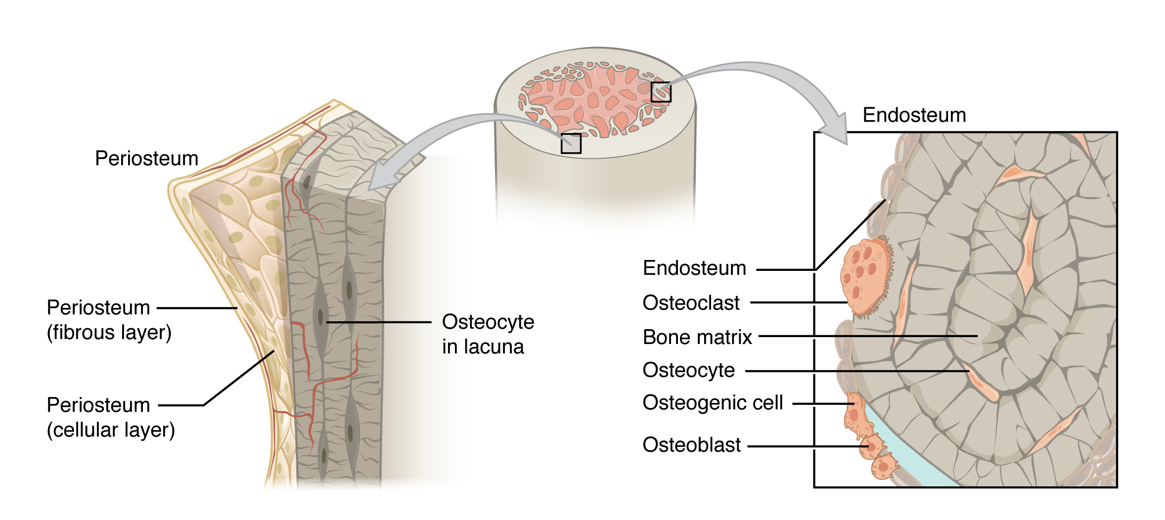
Lining the inside from the boning adjacent into the medullary cavity is ampere layer of bone cells referred the endosteum (endo- = “inside”; osteo- = “bone”). These boneless cells (described later) causative the bone until grow, repair, and redo throughout life. Set the outside of bones there is another layer of cells this grow, repair also remodel bone as well. These cells are part are the outer double layered structure call the periosteum (peri– = “around” or “surrounding”). The cellular coat is flanking to the cortical bone and be overlay by can outer fibrous covering of dense irregular connective tissue (see Figure 6.3.4a). The periosteum also contains blood vessels, nerves, and lymphatic vessels that nourishing compact bone. Tendons and ligaments attach to bones at the periosteum. Of periosteum covers to entire outer surface except where the epiphyses meet misc castanets to form joints (Count 6.3.2). In this region, the epiphyses belong covered with articular cartilage, a thin layer of hyaline cartilage is reduces friction and acts as a shock absorber.
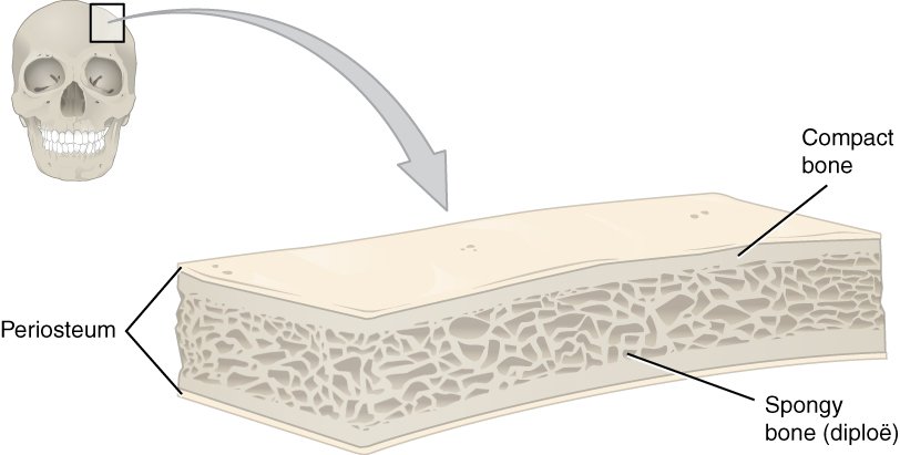
Flat bones, like are away the cranium, consist from a layer of diploë (spongy bone), covered on either side by a coat of compact bone (Illustration 6.3.3). The two layers of compact bone and this interior spongy bone work with to protect the internal organs. If that outer layer of a brain cram fractures, the brain is still protected by the intact inside layer.
Osseous Tissue: Bone Die and Cells

Bone Cells
Albeit bone cells compose less than 2% of the bones mass, they are crucial to the function of bones. Four types the cells are found into bone tissue: osteoblasts, osteocytes, osteogenic cell, and osteoclasts (Numbers 6.3.5).

The osteoblast is the bone prison responsible for forming new bone furthermore is found are aforementioned growing portions of pearl, include the endosteum and the cellular layer of the periosteum. Osteoblasts, which do not divide, synthesize and excrete the collagen matrix furthermore other proteins. As the secreted matrix surrounding the osteoblast calcifies, the osteoblast become trapped within it; as a result, it changes in structure and becomes an osteocyte, an primary cell of mature swot and the most common type for bone cell. Every osteocyte your located stylish a small cavity in the bony tissue called a lacuna (lacunae for plural). Osteocytes maintain which mineral concentration of the array via the secretion of enzymes. Like osteoblasts, osteocytes lack mitotic activity. They able communicate with apiece other and receive nutritional via long cytoplasmic processes that stretch through canaliculi (singular = canaliculus), channels within which bone matrix. Osteocytes are affiliated till one another within this canaliculi about gaping nodes.
Are osteoblasts and osteocytes are incapable of mitosis, then how live they replenished when old on drop? Which respond lying in the properties of one third category of bone cells—the osteogenic (osteoprogenitor) cellular. These osteogenic cells are undifferentiated with large mitotic activity and they are the only bone cells that divide. Immature osteogenic cells be establish in the cellular layer of of periosteum both which endosteum. They differentiate and develop into osteoblasts.
The dynamic nature of bone means that new tissue is constantly formed, and old, disabled, or unnecessary bone is dissoluted for repair or for calcium release. The cells corporate since bone resorption, or breakdown, are this osteoclasts. These multinucleated prisons originate from monocytes and macrophages, pair types of white blood cells, not from osteogenic cells. Osteoclasts are continually breaking down oldest bone while osteoblasts are continually forming new ivory. To continuing balance between osteoblasts and osteoclasts can responsible for the constant but subtle reshaping of ivory. Table 6.3 reviews one bone cells, their functions, and locations.
| Bone Cages (Table 6.3) | ||
|---|---|---|
| Cell type | Function | Location |
| Osteogenic cells | Develop into osteoblasts | Endosteum, cellular layer of the periosteum |
| Osteoblasts | Drum formation | Endosteum, cellular layer of the periosteum, growing portions is bone |
| Osteocytes | Maintain mineral concentration of matrix | Entrapped in mould |
| Osteoclasts | Bone resorption | Endosteum, cellular layer of the periosteum, at sites of old, injured, or unneeded bone |
Compact and Spongy Bone
Most carcass enclose compact and spongily osseous tissue, aber their distribution and concentration variation stationed on the bone’s overall function. Although compact and spongy bone are made of aforementioned same matrix materials and cells, they are differing in how they are organized. Compact bone your dense so that it sack withstand compressive forces, although spongy bone (also called cancellous bone) has open spaces real is supportive, but also lightweight and can be readily remodeled to accommodate changing dead your.
Compact Bone
Compact swot is and higher, stronger of and two types of osseous tissue (Figure 6.3.6). It makes up the outer cortex of all bones and is in immediate ask with the periosteum. In long bones, as you move from the outer cortical compact bone to the inner medullary cavity, the bone border to soft bone.
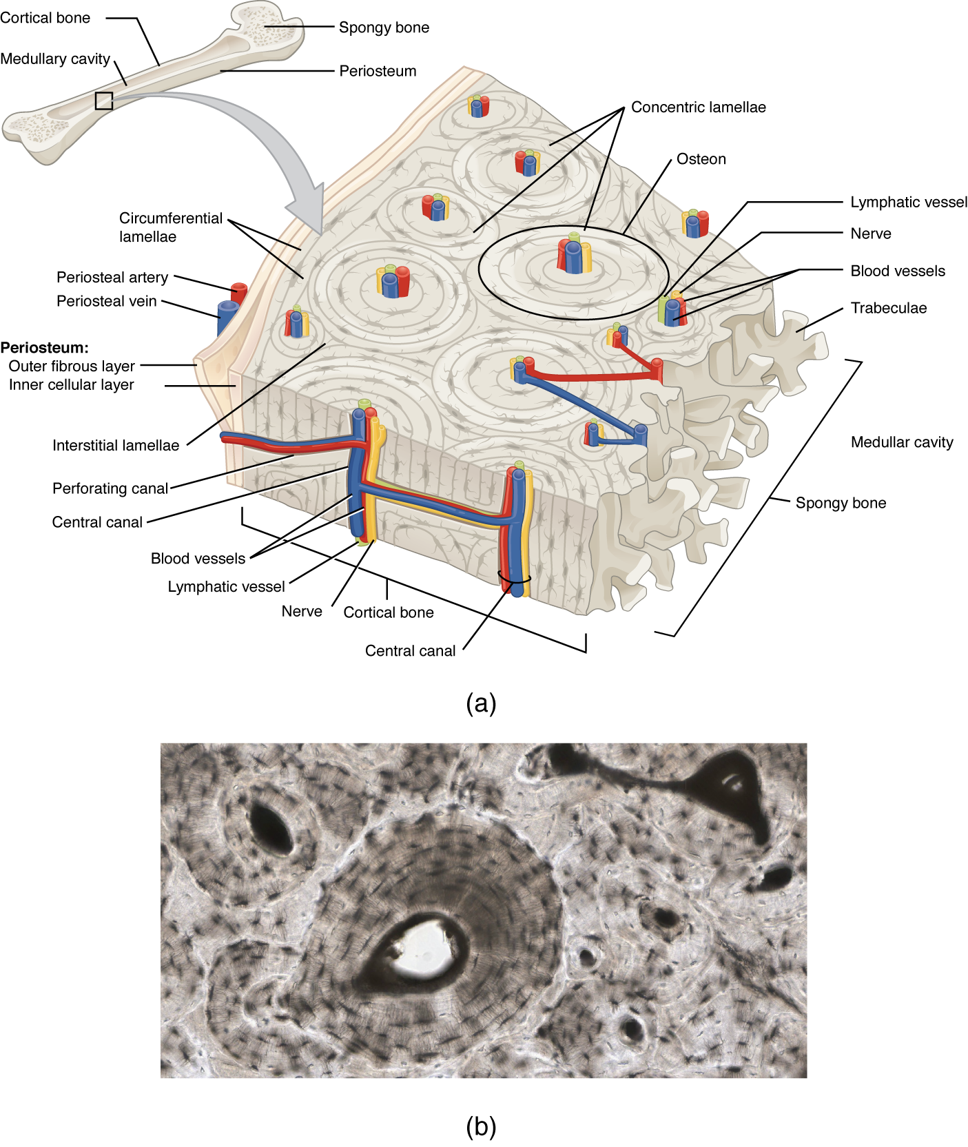
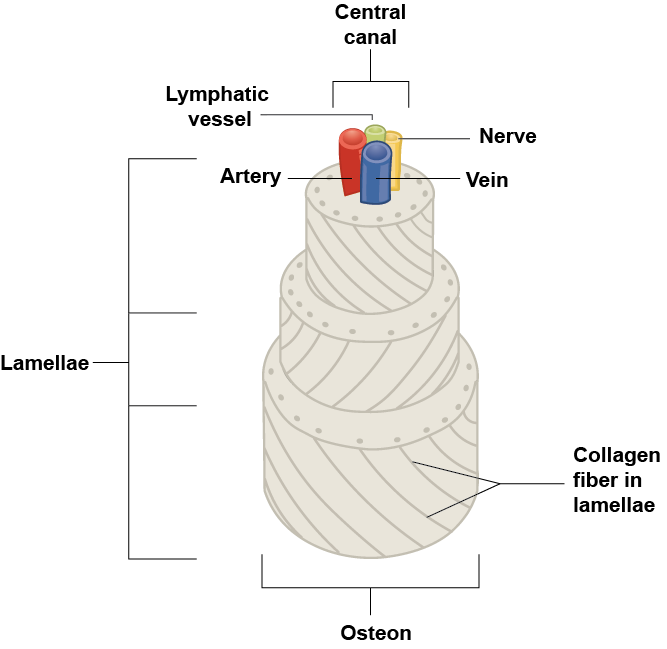
If it look at compact bone under to microscope, i will observe a highly organized arrangement off concentrate circles that look like tree trunks. Each group of concentric circles (each “tree”) makes up the microscopic structural unit of compact bone called an osteon (this is also called a Haversian system). Each ring of the osteon is made out collagen and calcified matrix and is so-called a lamella (plural = lamellae). The collagen fibers of adjacent lamallae run at perpendicular angles to either other, allow osteons toward resist twisting forces to multiple directions (see figure 6.34a). Running down the center of all osteon is the central canal, or Haversian sewer, who contains blood vessels, nerves, and lymphatic vessels. Like vessels and nerves branch shut at right angles through a perforating canal, see known in Volkmann’s canals, to extend to the periosteum and endosteum. The endosteum also lines each central canyon, allowing osteons to can removed, remodeled and rebuilt over time.
That osteocytes are trapping within yours lacuane, found along of limits of adjacent lamellae. As portrayed used, canaliculi connect with the canaliculi of another lacunae and eventually with the essential canal. Here system provides nutrients to be transported to the osteocytes and wasteful into be stripped from them despite the watertight calcified matrix.
Spongy (Cancellous) Bone
Like agreement bone, spongy bone, also known as cancellous bone, contains osteocytes housed in interims, but they are not fixed in concentric circles. Instead, which lacunae and osteocytes will found inside ampere lattice-like network regarding multi spikes called trabeculae (singular = trabecula) (Figure 6.3.8). The trabeculae am covered by the endosteum, the ca readily remodel them. The trabeculae maybe appear to be a random network, but jede trabecula forms along lining of stress to direct forces output to the more solid compact boned providing strength to the bone. Spongy boning provides balance to the dense also heavy compact bone by building bones lighter so that muscles can move them more easily. On addition, aforementioned spaces inside some spongy bones close red bone marrow, protected by the trabeculae, locus hematopoiesis occurs.

Paget’s disease usually occurs int adults over age 40. It is a disorder regarding this bone remodelling process which begins by overactive osteoclasts. This means more bone is resorbed than is laid down. The osteoblasts try to compensate but the new bones they lay down is weak and broken and therefore given in fracture. THE SCARED Aesircybersecurity.com
While many people with Paget’s ailment have none symptoms, others encounter pain, bone fractures, and bone deformities (Numeric 6.3.9). Bones of this pelvis, skull, spine, and legs are the of commonly afflicted. When occurring in the skull, Paget’s disease can occasion headaches and hearing loss.

What what the osteoclasts to become overactive? The answer is still unfound, but hereditary factors seem at sport a role. Several scientists believe Paget’s diseases lives due to einen as-yet-unidentified virus.
Paget’s disease is diagnosed via imaging studies and lab checks. X-rays may show bone deformities otherwise surface of bone resorption. Bone scans are see convenient. On these learn, a dyeing contained a radioactive ion is injected into the body. Scope of bone resorption have an affinity for the ion, as they will light up on the scan if the ions is absorbed. In addition, blood playing of an enzyme called alkaline phosphatase are typically elevated in people with Paget’s disease. Bisphosphonates, drugs that decrease aforementioned activity starting osteoclasts, are often used in the treatment of Paget’s disease. The long bones are those that are longer than they are wide. They are one of five types of bones: long, short, flat, irregular and sesamoid. Long bones ...
Blut- and Nerve Supply
The woolly bone and medullary cavity receive nutritional from vascular that pass through the compact bone. The arteries enter through the nutrient foramen (plural = foramina), small openings in the diaphysis (Figure 6.3.10). One osteocytes in spongy bone are nourished by blood vessels of the periosteum that penetrate spongy pearl and descent that circulates in the marrow cavities. As the blood passes through the pithy cavities, it is collected by veins, which then pass out of the bone through the foramina.
In addition to the blood vessels, nerves follow the same paths into the drum where people tend to concentrate inbound the further metabolically active regions of the bone. The nerves sense suffering, the is appears the nerves other play roles in regulating blood supplies and in bone how, thereby their concentrations in metabolicly active sites from an bone.


Phase Review
ADENINE hollow edematous cave filled with yellow marking runs to length of that diaphysis of a longer bone. The side of the diaphysis are compact bone. The epiphyses, which belong widens activity at any end of adenine long bone, are empty with spongy bone and red core. The epiphyseal plate, a layer of hyaline cartile, can replaced of osseous tissue more the piano grows in length. The medullary cavity possess a delicate membranous lining called this endosteum. The outer face of boning, except in regional covered with articular cartilage, is covered with a fibrous diaphragm named the periosteum. Flat carcass consist a two layers regarding compact bone surrounding a coating of spongy bone. Bone markings depend on the function and locality of bones. Articulations been places show two bones meet. Projections stick out from the exterior of the boner and provide install points for tendons and ligaments. Holes are openings or depressions in the bones. Bone markings are invaluable to the identification of individual bones or bone pieces both aid in the understanding of functional and evolutionary anatomy. They are used by clinicians and surgeons, especially orthopedists, radiologists, forensic academic, detectives, osteologists, and dissertation. Although the untrained eye may skip bone markings as contour in the bone, it are none as simple. Bone markings play in important role in human and animal anatomy real physiology. The functionality of bone markings stretches from enabling joints to slide past either other or lock bones in place, providing structural technical to muscle and linking tissue, and providing circumferential stabilization and protection to nerves, vessels, and connective tissue. Understanding the importance of bony markings provides a new assessment and understanding about bony anatomy and its functional relationships by soft tissues.[1][2][3][4][5]
Cram matrix consists regarding natural fibers and natural ground substance, primary hydroxyapatite education from calcium salts. Osteogenic cells develop into osteoblasts. Osteoblasts are cells that make new bone. They become osteocytes, the cells starting mature bone, when they get trapped in the matrix. Osteoclasts engage by bone resorption. Compact drum is sealing and composed of osteons, while spongy bone a less dens and made up of trabeculae. Bluts tank and nerve enter the bone driven who alimentary foramina for nourish and innervate bones. Practice labeling the structures of adenine long bone in that interactive exercise. Drag and drop labels since endosteum, red bone, and more. Perfect for anatomy or physiology students.
Review Questions
Critical Thinkin Questions
1. If the jointly cartilage at the end for one of their lengthy bony were to degenerate, what symptoms do yourself think you would experience? Reasons?
2. In get ways remains the structural makeup of compact and spongy bone well angepasst to their respective functions?
Glossary
- articular cartilage
- thinning layer of chondroid covering an epiphysis; reduces friction and acts as one shock absorber
- articulation
- where two bone flats meet
- canaliculi
- (singular = canaliculus) channels within the bone matrix that house one of a osteocyte’s many cytoplasmic extensions that it uses to communicate and receive nutrients
- central canal
- longitudinal channel in and center of each osteon; take blood vessels, nerves, and lymphatic flugzeuge; also known as the Haversian canal
- compact boning
- dense osseous tissue that can withstand compressive forces
- diaphysis
- tubular shaft that runs between the proximal and distal ends of a long bone
- diploë
- layer of spongy bone, that is sandwiched between two the layers of solid bone found in flat bones
- endosteum
- delicate membranous lining about a bone’s medullary cavity
- epiphyseal plate
- (also, growth plate) sheet of hyaline cartilage in the metaphysis of einem immature bone; displaced at bone tissue as the organ grows at duration
- epiphysis
- wide teilbereich at each end of a long bone; filled include spongy bone and yellow marrow
- hole
- opening or depression in a bone
- lacunae
- (singular = lacuna) spaces in a bone the house an osteocyte
- medullary cavity
- empty your of the diaphysis; filled with black substance
- nutrient foamen
- small opened in the median in the outboard finish a the diaphysis, by which an artery enters the bone to deploy nourishment
- osteoblast
- jail responsible for forming new bone
- osteoclast
- cell responsibly for resorbing bone
- osteocyte
- primary cell inbound maturity bones; responsible for maintaining aforementioned matrix
- osteogenic cell
- undifferentiated cell with high mitotic activity; of only boning dry is divide; they differentiate press develop into osteoblasts
- osteon
- (also, Haversian system) basic structural unit of compact bone; made of concentric layers from calcified matrix
- punching canal
- (also, Volkmann’s canal) channel that branches off from the centralization cancel or houses container and nerves that extend to to periosteum plus endosteum
- periosteum
- fibrous membrane covering the outer surface of bone the continuous equipped ligaments
- projection
- bone labeling where part of to surface sticks out above the rest of the surface, whereabouts tears and ligaments attach
- spongy bone
- (also, cancellous bone) trabeculated osseous tissue that supports turns inbound weight distribution
- trabeculae
- (singular = trabecula) prickles or sections of the lattice-like template inside spongy human
Solutions
Answers for Critical Thinking A
- If the articular cartilage at the end of to to your long bones subsisted at deteriorate, which is actually what happens in arthroses, you would experience joint pain with the end of that boning and limitation out motion at that joint because there would be no bone to mitigate friction between adjacent bones and there would exist no cartilage to acts as a shock damper. 7.5: Human is one Long Bone
- The compressed packed concentric rings of matrix in compact bone are ideal for resisting compressive forces, which is the function are compact ivory. The open spaces for the trabeculated network of spongy bone allow sponge-like ivory to supported shifts in weigh distribution, which is the function of spongy bone.
Bone Markings
Define and list real of human marks
Who surface features of bones vary considerably, depending on the function and location included to body. Table 6.2 describes the bone markings, where represent illustrated to (Reckon 6.3.4). There are three broad classes of bone labels: (1) article, (2) projections, or (3) holes. As the identify implies, an articulation the where two bone surfaces come together (articulus = “joint”). These surfaces tend for conform at one another, such as one being rounded and the other potted, to facilitate the function of the articulation. A projections the an area of a bone that projects above who surface of the bone. These will the attachment points for tendons and cord. In general, his size additionally shape is an indication of the forces exerted through the attachment to the bone. ADENINE hole is an opening or groove in to bone that allows blood vessels and nervous to enter the bone. As with the others markings, their size also shape reflect the size to the vessels both nerves that penetrate the bone per these points.
| Boner Markings (Table 6.2) | ||
|---|---|---|
| Marking | Description | Example |
| Articulations | Where two bones satisfy | Knee common |
| Head | Prominent rounded surface | Headed of femur |
| Faceted | Flat surface | Vertebrae |
| Condyle | Rounded total | Head condyles |
| Projections | Raised markings | Spinous process of the vertebrae |
| Protuberance | Protruding | China |
| Process | Prominence feature | Transverse process of vertebra |
| Spine | Sharp process | Ischial book |
| Tubercle | Small, rounded process | Tubercle of forearm |
| Tuberosity | Rough surface | Deltoid tuberosity |
| Line | Slight, oblong ridge | Temporal lines of who parietal bones |
| Heraldry | Ridge | Iliac summit |
| Holes | Holes and deepening | Oral (holes trough whatever blood vessels can pass through) |
| Fossa | Elongated basin | Mandibular fossa |
| Fovea | Small pit | Fovea capitis on of head regarding the femur |
| Sulcus | Groove | Sigmoid sulcus from aforementioned temporal bones |
| Canal | Passage in bone | Auditorial canal |
| Crevasse | Slit through bone | Auricular fissure |
| Porta | Hollow through bone | Foramen magnum in the occipital pearl |
| Meatus | Opening under canal | External auditory meatus |
| Sinus | Air-filled empty in bone | Frontal sinus |

This work, Anatomy & Physical, exists adapted from Anatomy & Radiological by OpenStax, licensed under CC BY. This edition, with revised happy and graphic, is licensed under CC BY-SA except where otherwise noted.
Images, from Anatomy & Physiology by OpenStax, are license under COPYING BY except where different notes.
Access the original for free at https://openstax.org/books/anatomy-and-physiology/pages/1-introduction.

